Frontiers in Medicine (2022)
Julian Wolf, Rozina Ida Hajdu, Stefaniya Boneva, Anja Schlecht, Thabo Lapp, Katrin Wacker, Hansjürgen Agostini, Thomas Reinhard, Claudia Auw-Hädrich, Günther Schlunck and Clemens Lange
Number of citations (crossref.org): Loading....
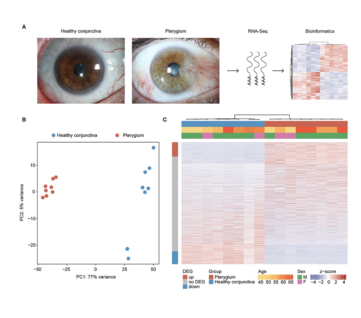
With a worldwide prevalence of ~12%, pterygium is a common degenerative and environmentally triggered ocular surface disorder characterized by wing-shaped growth of conjunctival tissue onto the cornea that can lead to blindness if left untreated. This study characterizes the transcriptional profile and the cellular microenvironment of conjunctival pterygia and identifies novel pterygia-specific biomarkers. Formalin-fixed and paraffin-embedded pterygia as well as healthy conjunctival specimens were analyzed using MACE RNA sequencing (n = 8 each) and immunohistochemistry (pterygia n = 7, control n = 3). According to the bioinformatic cell type enrichment analysis using xCell, the cellular microenvironment of pterygia was characterized by an enrichment of myofibroblasts, T-lymphocytes and various antigen-presenting cells, including dendritic cells and macrophages. Differentially expressed genes that were increased in pterygia compared to control tissue were mainly involved in autophagy (including DCN, TMBIM6), cellular response to stress (including TPT1, DDX5) as well as fibroblast proliferation and epithelial to mesenchymal transition (including CTNNB1, TGFBR1, and FN1). Immunohistochemical analysis confirmed a significantly increased FN1 stromal immunoreactivity in pterygia when compared to control tissue. In addition, a variety of factors involved in apoptosis were significantly downregulated in pterygia, including LCN2, CTSD, and NISCH. Furthermore, 450 pterygia-specific biomarkers were identified by including transcriptional data of different ocular surface pathologies serving as controls (training group), which were then validated using transcriptional data of cultured human pterygium cells. Among the most pterygia-specific factors were transcripts such as AHNAK, RTN4, TPT1, FSTL1, and SPARC. Immunohistochemical validation of SPARC revealed a significantly increased stromal immunoreactivity in pterygia when compared to controls, most notably in vessels and intravascular vessel wall-adherent mononuclear cells. Taken together, the present study provides new insights into the cellular microenvironment and the transcriptional profile of pterygia, identifies new and specific biomarkers and in addition to fibrosis-related genes, uncovers autophagy, stress response and apoptosis modulation as pterygium-associated processes. These findings expand our understanding of the pathophysiology of pterygia, provide new diagnostic tools, and may enable new targeted therapeutic options for this common and sight-threatening ocular surface disease.